VQ Scan: Understanding the Purpose, Procedure, and Interpretation
A VQ scan, a nuclear medicine imaging test, plays a crucial role in assessing lung health and diagnosing pulmonary conditions. In this comprehensive guide, we’ll delve into the fundamentals of the VQ scan, its significance, and the process involved.

What is a VQ Scan?
A VQ scan, short for ventilation-perfusion scan, is a specialized imaging technique used to evaluate lung function and blood flow. It’s particularly valuable in identifying conditions affecting lung ventilation and perfusion, such as blood clots, embolisms, and respiratory disorders. The scan combines two essential aspects: assessing how well air reaches the lungs and how well blood circulates within the lungs.
When is a VQ Scan Ordered?
A VQ scan is often recommended when healthcare providers suspect pulmonary abnormalities or suspect a blood clot in the lungs. Common symptoms that may warrant a VQ scan include shortness of breath, chest pain, and unexplained coughing. The scan is especially useful in diagnosing conditions like pulmonary embolism, chronic obstructive pulmonary disease (COPD), and lung cancer.
How is a VQ Scan Performed?
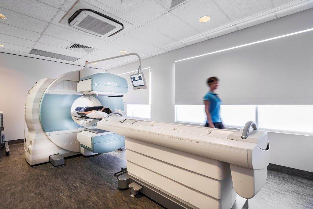
A VQ scan involves two essential components: ventilation and perfusion. During the ventilation phase, you’ll inhale a slightly radioactive gas through a mask. This gas allows technicians to capture images of the air distribution in your lungs. In the perfusion phase, a small amount of a radioactive dye is injected into a vein, usually in your arm. This dye travels through your bloodstream and highlights areas with good and poor blood flow in your lungs. Specialized imaging equipment, such as a gamma camera, is used to capture these images.
What to Expect During the Procedure?
Undergoing a VQ scan is a non-invasive and painless process. During the ventilation phase, you’ll be asked to breathe normally while wearing a mask that delivers the radioactive gas. During the perfusion phase, the dye injection might cause a temporary sensation of warmth. It’s important to remain still during the imaging process to ensure accurate results. The procedure typically takes around 1 to 2 hours to complete.
Interpreting VQ Scan Results
After the scan, a radiologist will carefully analyze the collected images. A key aspect of interpretation involves comparing the ventilation and perfusion images. Matching or mismatching areas provide valuable insights into lung function. A mismatch, where there’s good ventilation but poor blood flow (or vice versa), could indicate conditions like pulmonary embolism or other lung disorders.
Benefits and Limitations of VQ Scans
VQ scans offer several advantages. They provide valuable diagnostic information without the need for invasive procedures. Additionally, they expose patients to a relatively low dose of radiation. However, it’s important to note that VQ scans have limitations. Some factors, like obesity or pre-existing lung conditions, can affect the accuracy of results. Your healthcare provider will weigh these factors when interpreting the findings.
Preparing for a VQ Scan
To ensure accurate results, there are a few preparatory steps to follow before undergoing a VQ scan. You may be asked to avoid certain medications that could influence blood flow or interfere with the radioactive dye. It’s important to inform your healthcare provider about any allergies, previous surgeries, or medical conditions you have. Additionally, if you’re pregnant or breastfeeding, let your doctor know, as radiation exposure can potentially affect the fetus or infant.
Potential Risks and Safety Measures
While a VQ scan exposes you to a small amount of radiation, the associated risk is minimal. The benefits of accurate diagnosis often outweigh the potential risks. To ensure safety, the healthcare team takes precautions to minimize radiation exposure. If you have concerns about radiation, discussing them with your doctor can help alleviate any worries.
Alternatives to VQ Scans
In some cases, alternative imaging methods may be considered based on your medical history and the specific condition being investigated. CT scans and pulmonary angiograms are among the alternatives. Your healthcare provider will determine the most suitable imaging approach for your unique situation.
Frequently Asked Questions (FAQs) About VQ Scan
Q1: What is a VQ scan?
A1: A VQ scan, also known as a ventilation-perfusion scan, is a medical imaging test used to assess lung ventilation and blood circulation.
Q2: Why is a VQ scan performed?
A2: A VQ scan helps diagnose lung conditions such as pulmonary embolism, blood clot-related issues, and lung disorders by evaluating air distribution and blood flow.
Q3: What does the procedure involve?
A3: The procedure includes inhaling a slightly radioactive gas to assess ventilation and receiving a radioactive dye injection to evaluate perfusion. Specialized imaging equipment captures lung images.
Q4: Is the procedure painful?
A4: No, a VQ scan is generally non-invasive and painless. The dye injection may cause a temporary sensation of warmth.
Q5: How long does the procedure take?
A5: The procedure typically takes around 1 to 2 hours to complete, including both the ventilation and perfusion phases.
Q6: Are there any risks associated with radiation exposure?
A6: The radiation exposure during a VQ scan is minimal and generally considered safe. The benefits of accurate diagnosis usually outweigh the risks.
Q7: Can anyone undergo a VQ scan?
A7: Most individuals can undergo a VQ scan, but certain factors like pregnancy and allergies should be discussed with your healthcare provider before the procedure.
Q8: Are there alternatives to VQ scans?
A8: Yes, alternatives include CT scans and pulmonary angiograms. The choice depends on your medical history and the specific condition being investigated.
Q9: What do mismatched areas in the results indicate?
A9: Mismatched areas, where ventilation and perfusion do not match, could indicate conditions like pulmonary embolism or other lung disorders.
Q10: How can I prepare for a VQ scan?
A10: Preparing may involve avoiding certain medications, informing your doctor about allergies or medical history, and discussing concerns about radiation exposure.
Conclusion
VQ scans play a vital role in diagnosing and assessing lung conditions, providing valuable insights into lung ventilation and blood flow. With their non-invasive nature and ability to provide important diagnostic information, VQ scans empower healthcare professionals to make accurate treatment decisions. If you’re recommended for a VQ scan, understanding the procedure and its significance can contribute to a well-informed and proactive approach to your lung health.


