Decoding Knee MRI: A Comprehensive Guide to Understanding and Interpreting Results
An MRI (Magnetic Resonance Imaging) of the knee is a crucial diagnostic tool for evaluating and diagnosing various knee injuries and conditions. This comprehensive guide aims to provide a step-by-step breakdown of knee MRI, interpretation, and common FAQs to help you understand and interpret the results effectively.

Understanding Knee MRI Basics
What is a Knee MRI?
A knee MRI is a non-invasive medical imaging technique that uses powerful magnets and radio waves to capture detailed images of the knee joint. Unlike X-rays or CT scans, MRI provides a more comprehensive view of the knee’s soft tissues, including ligaments, tendons, cartilage, and muscles. It plays a crucial role in diagnosing and evaluating knee conditions, such as ligament tears, meniscal injuries, and osteoarthritis.
How Does Knee MRI Work?
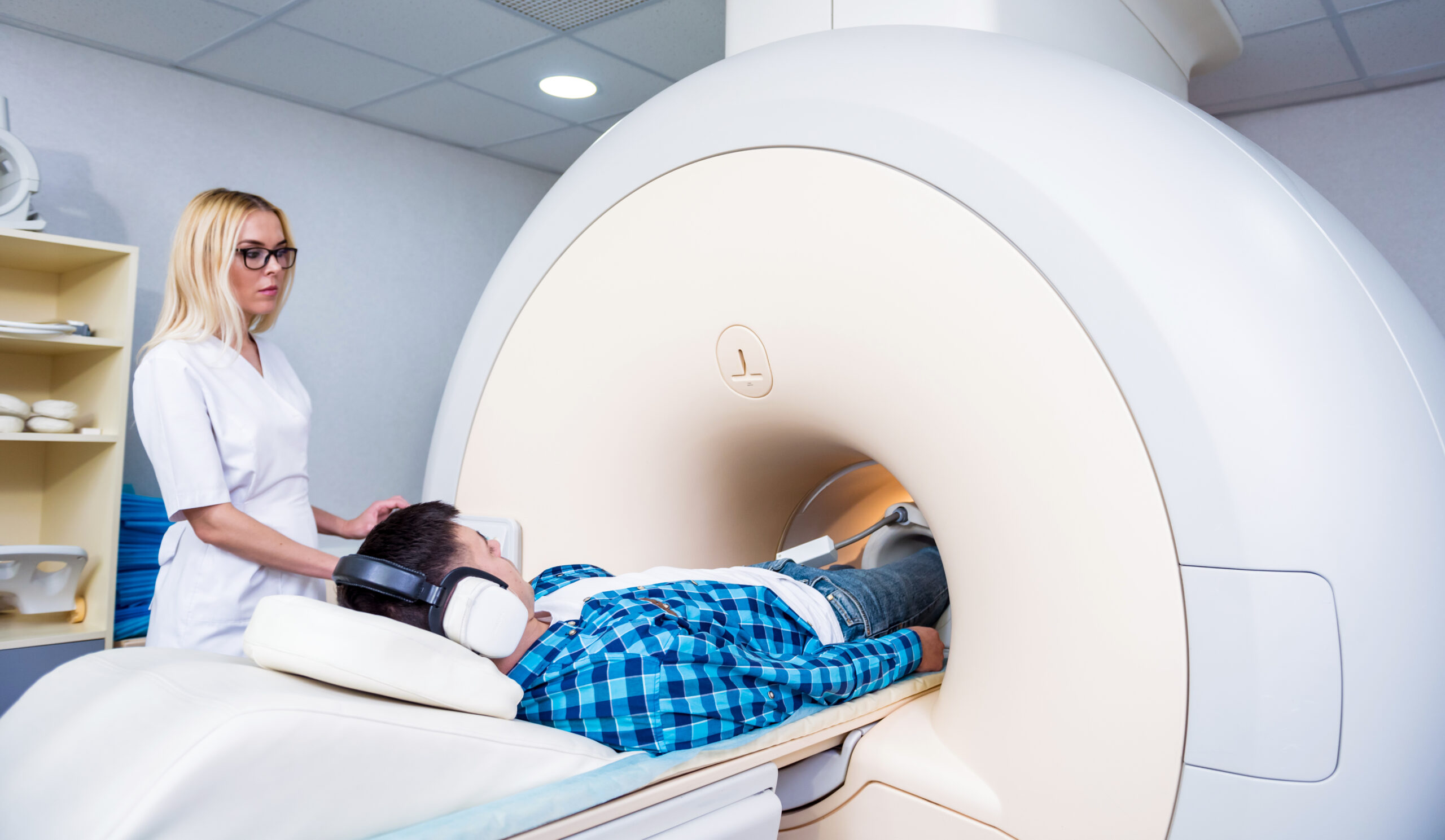
Knee MRI utilizes magnetic fields and radio waves to create detailed images of the knee. The patient lies down on a table that slides into the MRI machine, which contains a strong magnet. The machine emits radio waves that cause the body’s atoms to produce signals. These signals are captured by the machine and processed into high-resolution images by a computer. The entire scanning process typically takes around 30-60 minutes.
Preparing for a Knee MRI
Before a knee MRI, it is important to follow certain instructions. You may be asked to remove clothing and jewelry that contain metal, as the magnetic field can interfere with the imaging process. You should also inform the healthcare provider if you have any metal implants or devices in your body. If you experience claustrophobia or anxiety, you can discuss your concerns with the healthcare provider, who may offer options to help you feel more comfortable during the procedure.
What to Expect During a Knee MRI
During a knee MRI, you will be positioned on the table and given earplugs or headphones to minimize the noise generated by the machine. It is important to remain still and follow the instructions provided by the MRI technician. The machine will make loud knocking or buzzing sounds during the scanning process, but it is normal. If you experience any discomfort or need assistance, there will be a communication system to communicate with the technician. The entire procedure is painless, and you will be able to resume your normal activities afterward.
Interpreting Knee MRI Results
Components of a Knee MRI Report
A knee MRI report typically consists of several sections that provide important information about the imaging findings. These sections may include:
Images: The report may include images of the knee from different angles, allowing healthcare professionals to visualize the structures and abnormalities.
Impressions: This section highlights the radiologist’s observations and interpretations of the images, including any abnormalities or significant findings.
Recommendations: The report may provide recommendations for further evaluation or treatment based on the MRI findings.
It is essential to consult with a radiologist or orthopedic specialist to interpret the MRI report accurately and understand the implications of the findings.
Common Knee MRI Findings
Knee MRI can reveal various common findings related to knee injuries and conditions. Some of the frequently encountered findings include:
Ligament Tears: MRI can identify tears or injuries to the anterior cruciate ligament (ACL), posterior cruciate ligament (PCL), medial collateral ligament (MCL), and lateral collateral ligament (LCL).
Meniscal Injuries: MRI can detect tears or degeneration of the menisci, which are the cartilage pads that cushion the knee joint.
Osteoarthritis: MRI can show signs of joint degeneration, such as cartilage loss, bone spurs, and inflammation, indicating osteoarthritis.
These are just a few examples, and the specific findings in an MRI report will depend on the individual case. The images provided in the report will help healthcare professionals visualize and assess the extent of the identified abnormalities.
Interpreting Abnormalities and Artifacts
While knee MRI is highly accurate, there are instances where abnormalities or artifacts may appear in the images. It is crucial to differentiate between true abnormalities and imaging artifacts. Artifacts can occur due to patient movement, metal objects, or other factors that can distort the images.
Experienced radiologists can distinguish between true abnormalities and artifacts by carefully analyzing the images and considering the clinical context. They may request additional imaging or take other factors into account to ensure an accurate interpretation of the MRI findings.
Correlating MRI Findings with Symptoms
While knee MRI provides valuable information about the internal structures of the knee, it is essential to correlate the MRI findings with the patient’s clinical symptoms. The presence of abnormalities in the MRI does not always indicate that they are the cause of the symptoms.
Healthcare professionals, including radiologists and orthopedic specialists, play a crucial role in interpreting the MRI findings in the context of the patient’s symptoms and medical history. They consider both the objective imaging findings and the subjective complaints to determine the relevance and severity of the identified abnormalities. This comprehensive approach ensures accurate diagnosis and appropriate treatment planning.
Frequently Asked Questions
What are the risks and safety considerations associated with knee MRI?
Knee MRI is considered a safe procedure with no known risks or side effects. Unlike X-rays or CT scans, MRI does not involve ionizing radiation. However, it is important to inform the healthcare provider about any metal implants or devices in your body, as they may be affected by the strong magnetic field. Additionally, individuals with claustrophobia or anxiety may experience discomfort during the procedure, but measures can be taken to help alleviate these concerns.
How long does a knee MRI take?
The duration of a knee MRI procedure typically ranges from 30 to 60 minutes. However, the actual scanning time may vary depending on the specific imaging sequences required and the patient’s cooperation. In some cases, contrast agents may be administered to enhance certain structures, which may slightly prolong the procedure.
Can I drive myself home after a knee MRI?
In most cases, you can drive yourself home after a knee MRI. The procedure does not typically involve sedation or anesthesia that would impair your ability to drive. However, if contrast agents are used, it is advisable to confirm with your healthcare provider whether any driving restrictions apply. If you feel uncomfortable driving after the procedure, it is always a good idea to arrange for alternative transportation.
Are there any alternatives to knee MRI for diagnosing knee conditions?
While knee MRI is highly effective in diagnosing knee conditions, there are alternative imaging techniques available. These include X-rays, CT scans, and ultrasound. Each method has its advantages and limitations.
Conclusion:
A knee MRI is a vital tool in diagnosing and evaluating knee injuries and conditions. This comprehensive guide has provided an overview of knee MRI basics, interpretation of results, and answered some common FAQs. It is essential to consult with healthcare professionals, such as radiologists or orthopedic specialists, for accurate interpretation and guidance based on your specific case. By understanding and decoding knee MRI results, you can play an active role in your healthcare journey and improve treatment outcomes.


