Understanding the Woods Lamp Examination
In the world of dermatology, the Woods lamp has become an indispensable tool for dermatologists and skincare professionals. This simple yet powerful device plays a significant role in skin analysis and the early detection of various skin conditions. In this article, we will dive deep into what a Woods lamp is, how it works, and its crucial applications in dermatological practices.

What is a Woods Lamp?
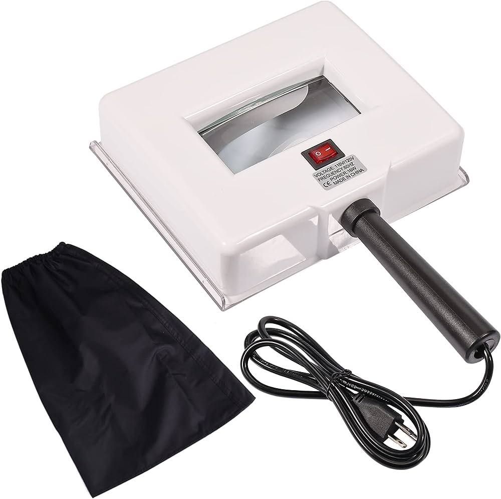
A Woods lamp, also known as a black light or ultraviolet (UV) lamp, is a handheld device that emits specific wavelengths of UV light. Named after its inventor, Robert Woods, this lamp is used extensively by dermatologists to observe the skin in a different light—literally. The Woods lamp emits UV light, which reveals hidden details and characteristics of the skin that are not visible to the naked eye under regular lighting.
Keywords:
Woods lamp, black light, ultraviolet lamp, UV light, skin analysis
LSI Keywords:
Dermatology tool, skin examination, UV light in dermatology, skin conditions detection
How Does a Woods Lamp Work?
The functioning of a Woods lamp revolves around the interaction between ultraviolet light and certain skin pigments and substances. When UV light is directed onto the skin, it causes various compounds to fluoresce, emitting different colors of light. This fluorescence enables dermatologists to identify specific substances within the skin layers, thereby aiding in the diagnosis of various skin conditions.
A Woods lamp examination involves the dermatologist closely observing the skin’s response to the UV light. Different colors and patterns that appear under the lamp’s illumination can provide valuable insights into the presence of skin disorders, infections, and irregularities.
Keywords: Ultraviolet light interaction, skin pigments fluorescence, skin disorders diagnosis
LSI Keywords: UV light impact on skin, skin pigments response, detecting skin infections
Clinical Applications of a Woods Lamp
The applications of a Woods lamp extend far beyond its seemingly simple design. Dermatologists leverage its unique abilities to detect and diagnose a range of skin conditions and concerns. Some common clinical applications include:
Fungal Infections:
Woods lamp can aid in identifying fungal infections like ringworm by highlighting the presence of fungal elements on the skin’s surface.
Bacterial Infections:
Certain bacterial infections, such as Porphyria cutanea tarda, exhibit distinct fluorescence under the UV light.
Vitiligo Detection:
In cases of vitiligo, where pigmentation loss occurs, the Woods lamp can help determine the extent of depigmentation.
Benefits and Limitations of the Woods Lamp
The Woods lamp offers several benefits that make it an essential tool in dermatology practices. However, like any diagnostic tool, it also comes with certain limitations. Let’s explore both aspects to gain a comprehensive understanding:
Benefits of the Woods Lamp
Early Detection:
The Woods lamp enables dermatologists to identify skin conditions in their early stages, allowing for prompt intervention and treatment.
Non-Invasive:
Unlike certain invasive procedures, a Woods lamp examination is non-invasive and painless for the patient.
Quick and Convenient:
The procedure is relatively quick and can be performed during routine dermatological examinations.
Enhanced Diagnosis:
It provides dermatologists with additional information that complements the visual examination of the skin.
Limitations of the Woods Lamp
Limited Accuracy:
While the Woods lamp can highlight certain conditions, it’s not a definitive diagnostic tool. Additional tests may be required for accurate diagnosis.
Skin Tone Variability:
Skin tones can influence the fluorescence patterns, making interpretation challenging for patients with darker skin.
Specific Conditions:
The lamp is most effective in diagnosing conditions related to pigmentation, fluorescence, and certain infections.
Common Skin Conditions Detected by a Woods Lamp
One of the most remarkable aspects of the Woods lamp is its ability to uncover hidden skin conditions that might not be immediately visible to the naked eye. Here are some common skin conditions that dermatologists can detect using this powerful tool:
Fungal Infections:
The Woods lamp can reveal the presence of fungal infections like tinea capitis (scalp ringworm) by detecting fluorescence in affected areas.
Pigmentation Irregularities:
Conditions like hypopigmentation and hyperpigmentation can be highlighted under the lamp’s UV light.
Bacterial Infections:
Certain bacterial infections, including Erythrasma, exhibit distinct fluorescence patterns, aiding in diagnosis.
Vitiligo Examination:
In vitiligo cases, the lamp helps visualize the extent of depigmentation, guiding treatment decisions.
Porphyria Detection:
Porphyria, a group of rare disorders, can be suspected through fluorescence patterns caused by accumulated porphyrins.
Preparing for a Woods Lamp Examination
If you’re scheduled for a Woods lamp examination, a few simple steps can help ensure accurate results. Here’s how to prepare for the procedure:
Clean Skin:
Arrive with clean and makeup-free skin. This allows the dermatologist to observe your skin’s natural state without any interference.
Remove Nail Polish:
If you have nail concerns, consider removing nail polish before the examination. Nails can exhibit fluorescence patterns under the UV light.
Discuss Concerns:
Communicate any skin concerns or symptoms you’re experiencing. This information helps the dermatologist focus on specific areas during the examination.
Interpreting the Woods Lamp Results
After the Woods lamp examination, your dermatologist will analyze the observed fluorescence patterns and colors. Here’s a general guide to interpreting the results:
Blue-Green Fluorescence:
Common in fungal infections like Microsporum canis (ringworm). The fluorescence indicates the presence of fungal elements.
White Fluorescence:
May suggest hypopigmentation or a fungal infection like tinea versicolor.
Purple-Red Fluorescence:
Could be indicative of porphyrin accumulation, associated with conditions like porphyria.
Lack of Fluorescence:
In certain cases, areas lacking fluorescence might indicate normal skin or different skin conditions.
Importance of Professional Examination
While the Woods lamp is a valuable tool, it’s crucial to remember that a professional’s expertise is necessary to accurately interpret results. Dermatologists undergo extensive training to analyze the subtle fluorescence patterns and diagnose conditions effectively.
Self-diagnosis solely based on Woods lamp results is discouraged, as other diagnostic methods and a holistic approach are often required for precise diagnosis and treatment recommendations.
FAQs About Woods Lamp Examination
Q: What is a Woods lamp examination?
A: A Woods lamp examination involves the use of a handheld device that emits UV light to observe skin fluorescence, aiding in the detection of various skin conditions.
Q: Is a Woods lamp examination painful?
A: No, a Woods lamp examination is non-invasive and painless. The UV light is simply directed onto the skin’s surface.
Q: How does a Woods lamp work?
A: A Woods lamp emits UV light that causes certain skin pigments and substances to fluoresce, revealing hidden details and characteristics of the skin.
Q: What skin conditions can a Woods lamp detect?
A: A Woods lamp can detect conditions like fungal infections (ringworm), bacterial infections, vitiligo, and pigmentation irregularities.
Q: Can a Woods lamp diagnose skin conditions definitively?
A: While it provides valuable insights, a Woods lamp is not a definitive diagnostic tool. Additional tests may be needed for accurate diagnosis.
Q: Are there limitations to the accuracy of a Woods lamp examination?
A: Yes, skin tone variability and specific conditions can influence fluorescence patterns, affecting interpretation.
Q: How can I prepare for a Woods lamp examination?
A: Arrive with clean, makeup-free skin, and consider removing nail polish. Communicate your skin concerns to the dermatologist.
Q: Can I self-diagnose using the results of a Woods lamp examination?
A: It’s not recommended. Professional interpretation by a trained dermatologist is crucial for accurate diagnosis and treatment.
Q: What do different fluorescence colors indicate?
A: Blue-green fluorescence can suggest fungal infections, white fluorescence may indicate hypopigmentation, and purple-red fluorescence could point to porphyrin accumulation.
Q: Is consulting a dermatologist necessary after a Woods lamp examination?
A: Absolutely. Dermatologists possess the expertise to interpret fluorescence patterns accurately and provide comprehensive diagnoses and treatment recommendations.
Conclusion
The Woods lamp has revolutionized the field of dermatology by providing dermatologists with a unique perspective on skin conditions. From fungal infections to pigmentation irregularities, its ability to reveal hidden details enhances the accuracy of diagnoses. However, it’s important to emphasize that the Woods lamp is just one piece of the diagnostic puzzle.


