Ophthalmoscopes: A Comprehensive Guide to Understanding and Usage
The world of ophthalmology relies heavily on advanced tools to ensure accurate diagnoses and effective treatments. Among these tools, the ophthalmoscope stands out as an invaluable device that allows medical professionals to peer into the intricate structures of the eye. In this comprehensive guide, we’ll delve into the multifaceted realm of ophthalmoscopes, shedding light on their history, types, importance in eye examinations, and more.
What is an Ophthalmoscope?
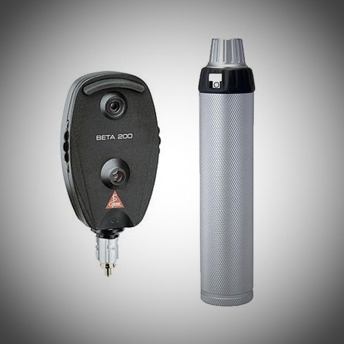
Imagine the human eye as a mesmerizing universe filled with details waiting to be unveiled. An ophthalmoscope acts as our telescope, revealing the mysteries within. It’s a handheld instrument that combines magnification and illumination to enable healthcare practitioners to inspect the retina, optic nerve, blood vessels, and other crucial components of the eye.

History and Evolution: From Rudimentary Tools to Modern Devices
The journey of the ophthalmoscope begins in the early 19th century when German physician Hermann von Helmholtz conceptualized the instrument. His creation marked the dawn of a new era in ophthalmology, allowing doctors to visualize the interior of the eye in ways previously deemed impossible. Over time, ophthalmoscopes have undergone remarkable transformations, evolving from simple handheld devices to sophisticated digital tools that integrate cutting-edge technologies.
Importance of Ophthalmoscopes in Eye Examination:
A thorough eye examination entails more than just testing visual acuity. Ophthalmoscopes play a pivotal role in diagnosing a spectrum of eye conditions, ranging from common issues to serious diseases. These include macular degeneration, diabetic retinopathy, glaucoma, and hypertensive retinopathy. By providing a direct view of the retina’s condition, ophthalmoscopes empower medical professionals to make accurate assessments, recommend appropriate interventions, and ultimately enhance patients’ ocular health.
Types of Ophthalmoscopes and Their Applications:
Ophthalmoscopes come in various types, each tailored to different examination needs. The direct ophthalmoscope is a basic handheld device used to inspect the fundus—the inner lining of the eye. Indirect ophthalmoscopes provide a wider field of view and are particularly useful for assessing peripheral retinal areas. For more detailed examinations, the slit-lamp ophthalmoscope offers enhanced magnification and illumination, making it indispensable in diagnosing intricate eye conditions.
Ophthalmoscopy Techniques and Best Practices:
Proper ophthalmoscopy technique requires skill and precision. Begin by dimming the lights to facilitate pupil dilation and patient comfort. Utilize the appropriate ophthalmoscope for the examination type. Position the patient comfortably and stabilize their head. Gently guide the ophthalmoscope’s light beam across the retina while focusing on the optic nerve head, blood vessels, and macula. Document findings accurately to track changes over time.
Common Eye Conditions Diagnosed with Ophthalmoscopes:
Ophthalmoscopy serves as a diagnostic gateway to various eye conditions. For instance, retinal detachment presents with the retina pulling away from its underlying tissue, often visible as a shadow or tear on the retina. Hypertensive retinopathy showcases characteristic changes in blood vessels due to high blood pressure. Optic nerve disorders such as glaucoma manifest as changes in the optic nerve head appearance. Early detection through ophthalmoscopy allows prompt intervention to preserve vision.
Integrating Ophthalmoscopy into Comprehensive Eye Care:
Collaboration between ophthalmologists and optometrists is essential for comprehensive eye care. Ophthalmoscopy supplements visual acuity tests, enabling practitioners to identify underlying issues that may impact vision. Regular eye examinations featuring ophthalmoscopy can lead to early detection of eye diseases and guide tailored treatment plans, emphasizing the significance of holistic eye care.
Technological Advancements in Ophthalmoscopes:
The digital age has propelled ophthalmoscope technology to unprecedented heights. Digital imaging capabilities allow for precise documentation and facilitate remote consultations. Smartphone-compatible ophthalmoscopes offer convenience, enabling quick snapshots of retinal conditions. Artificial Intelligence (AI) integration aids in automated analysis, potentially revolutionizing diagnostic accuracy and efficiency.
Training and Skill Development: Mastering the Art of Ophthalmoscopy:
Proficiency in ophthalmoscopy demands practice and education. Medical professionals can access workshops, seminars, and online resources to refine their skills. Hands-on training ensures accurate examination and interpretation of findings, guaranteeing optimal patient care and reliable diagnoses.
Frequently Asked Questions (FAQs) About Ophthalmoscopes
What is an ophthalmoscope and how does it work?
An ophthalmoscope is a medical device used to examine the interior structures of the eye, such as the retina and optic nerve. It combines magnification and illumination to provide a clear view of these structures.
What are the different types of ophthalmoscopes?
There are three main types of ophthalmoscopes: direct ophthalmoscopes, indirect ophthalmoscopes, and slit-lamp ophthalmoscopes. Each type offers distinct benefits for various examination purposes.
How is ophthalmoscopy performed?
Ophthalmoscopy involves dilating the patient’s pupils, dimming the lights, and using the ophthalmoscope to shine light into the eye while observing the reflection of the retina. This allows medical professionals to visualize its structures.
Why is ophthalmoscopy important in eye exams?
Ophthalmoscopy enables early detection of eye conditions like diabetic retinopathy, macular degeneration, and glaucoma. Detecting these conditions in their early stages improves the chances of successful treatment.
Are there any risks associated with ophthalmoscopy?
Ophthalmoscopy is a non-invasive procedure and generally safe. However, some patients may experience temporary discomfort from the bright light or eye drops used for pupil dilation.
How often should ophthalmoscopy be performed?
Regular eye exams, including ophthalmoscopy, are recommended annually for individuals with diabetes, high blood pressure, or a family history of eye diseases. Otherwise, it’s recommended every 2-4 years.
Can ophthalmoscopy detect systemic health issues?
Yes, ophthalmoscopy can sometimes reveal signs of systemic health conditions such as hypertension and diabetes by showing changes in the blood vessels of the retina.
Are there any advancements in ophthalmoscope technology?
Yes, modern ophthalmoscopes often have digital imaging capabilities, allowing for detailed documentation of the retina. Some can even integrate with smartphones for easy image sharing.
Is ophthalmoscopy uncomfortable for patients?
The procedure itself is not painful, but patients might experience temporary sensitivity to light due to pupil dilation. However, discomfort is usually minimal and short-lived.
Can ophthalmoscopy be performed on children?
Yes, ophthalmoscopy can be performed on children as part of routine eye exams. It’s important for monitoring eye health and detecting potential issues early.
Conclusion:
In the realm of eye care, ophthalmoscopes are the windows through which medical professionals gain profound insights into ocular health. From their humble origins to the current age of digital innovation, these devices have revolutionized diagnostics, treatment, and patient outcomes. As we continue to unlock new possibilities through advanced technologies, the art of ophthalmoscopy remains an integral part of safeguarding vision and enhancing overall well-being. Regular eye examinations that encompass ophthalmoscopy are a cornerstone of proactive healthcare, guiding us toward a future where sight is cherished and preserved.


